VATECH MENA |
All Rights Reserved | Privacy Policy

The PaX-i provides the most precise high-quality panoramic image by combining image processing and accumulated experience in dental imaging from VATECH. This will improve your diagnostic accuracy by increasing treatment planning and patient satisfaction.
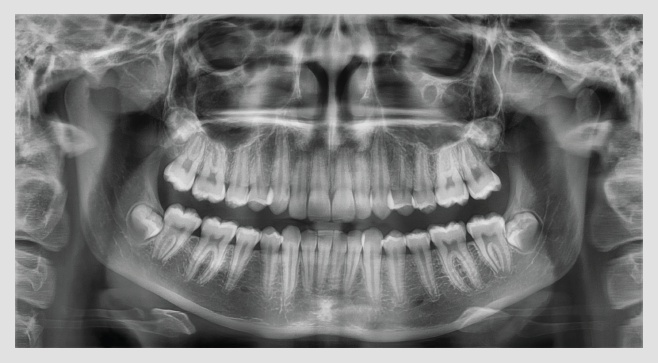
A clear and sharp panoramic image brings you better diagnostics.
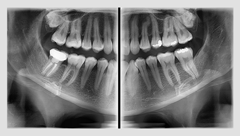
Advanced details, especially in the anterior and roots, can easily be viewed with the PaX-i.
These consistently high-quality images are the new standard of panoramic imaging.
The PaX-i has various capture modes to meet your diagnostic needs.
You can choose any capture mode based on your diagnostic needs.

| SELECTION | ARCH | EXAMINATION MODE |
|---|---|---|
| PANO EXAMINATION | Narrow / Normal / Wide / Child | Standard / Right / Front / Left |
| PANO EXAMINATION | Orthogonal | Orthogonal Standard / Right / Front / Left Bitewing Standard / Right / Front / Left |
| SPECIAL EXAMINATION | Normal | TMJ LAT Open / Close TMJ PA Open / Close Sinus LAT / PA |
EXTENDED DIAGNOSTIC VALUE FOR WIDE INSIGHT
The PaX-i provides optimal images exclusively designed for orthodontics.
There are two image sizes available (lateral and full lateral) that
allow you to choose your image size based on your diagnostic needs.
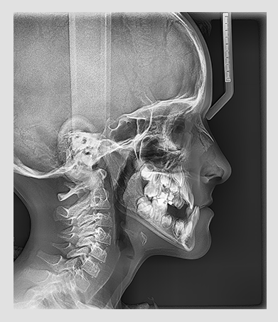
Provide specialized high quality images to suit
orthodontics and maxillofacial surgeries.
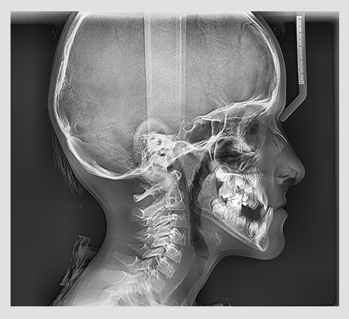
Full lateral image size is 30% wider and shows the
occipital area of the patient, which enables
comprehensive diagnosis.
| EXAMINATION PROGRAM | SCAN TIME | IMAGE SIZE |
|---|---|---|
| LATERAL | 12.9 sec | 21x23cm (8.3x9.1") |
| FULL LATERAL | 16.9 sec | 27x23cm (10.6x9.1") |
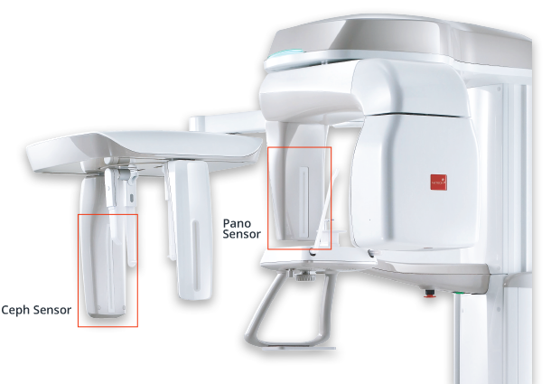
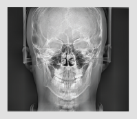
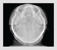
Superior image quality will be delivered using the a-Si TFT
sensors.Three different ceph image sizes reduce unnecessary x-ray dosage and scan the ideal area of cranial anatomy for your diagnosis and treatment planning.

| TYPE | PANO | CEPH (SCAN) | CEPH (ONE SHOT) |
|---|---|---|---|
| PAX-I | | | |
| PCH-2500 | | | |
| FUNCTION | SCAN TIME | FOCAL SPOT | TUBE VOLTAGE/CURRENT |
|---|---|---|---|
| Pano + Ceph | Pano : HD 13.5 sec/ Normal 10.1 sec Ceph : Scan 12.9 sec |
0.5 mm | 50-90 kVp/ 4-10 mA |
| SENSOR TYPE | PATIENT POSITION | WEIGHT |
|---|---|---|
| CMOS | standing / wheelchair accessble |
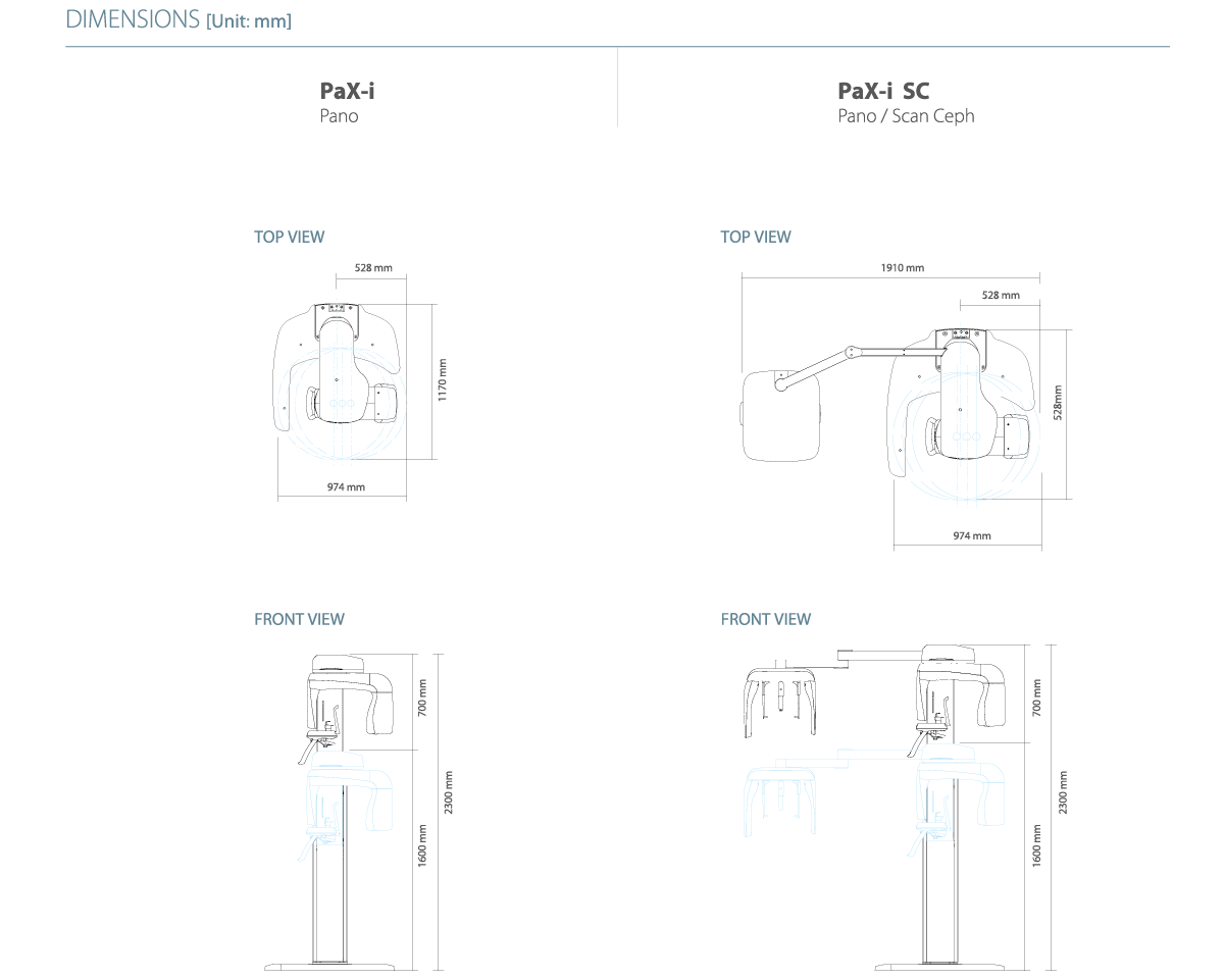
pax-i without base 90 kg pax-i sc without base 120 kg Base 50 kg |
* The specifications are subject to change without prior notice.