
VATECH MENA |
All Rights Reserved | Privacy Policy
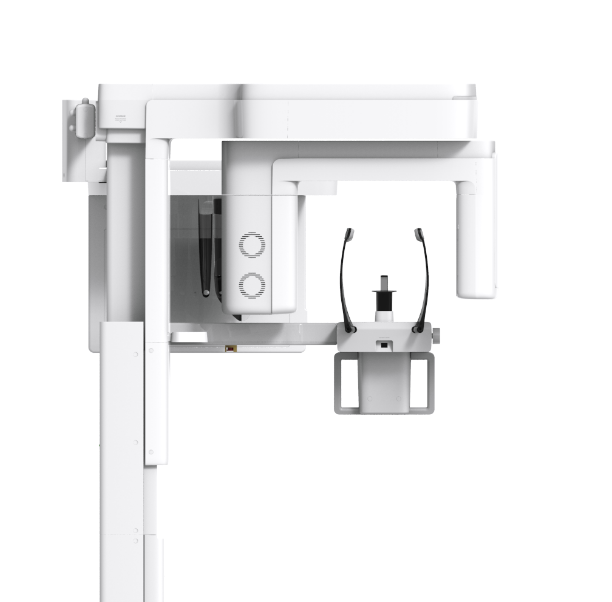
Green X Is an advanced 4in-1 digital x-ray Imaging system thet Incorporates Pano, Ceph {optional}, CBCT, and Model Scan. With VATECH’s extensive experience in the dental imaging field, the Green X provides high-quality Images with lower radiation by combining image processing.
This will improve your diagnostic accuracy and lead to Increased treatment planning end patient satisfaction.
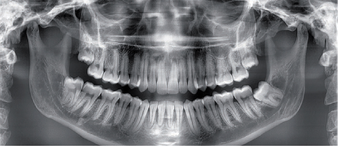
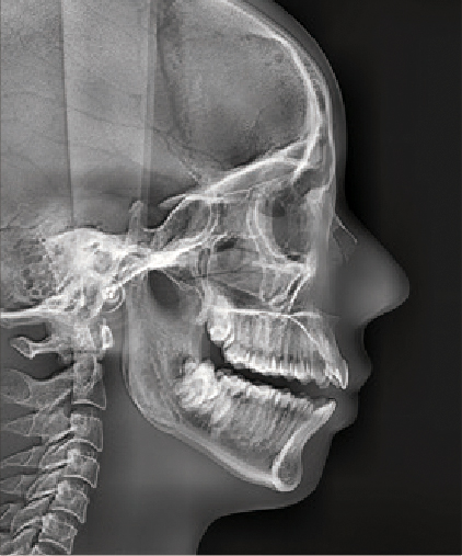


Due to its scan time, the Green X minimizes motion artifact and enables faster work flow. It produces superb diagnostic images, which will be a source of pride for any dental practice. Focusing on the highest quality of patient care, VATECH strives to improve the health and safety of your patients.


With its 4cm x 4cm volume mode and 49.5 micron voxel size, the Endo mode will optimize treatment of highly-focused regions of interest. It is ideal for endodontic use because the dentist is able to achieve an extraordinary image in a high-resolution voxel size.
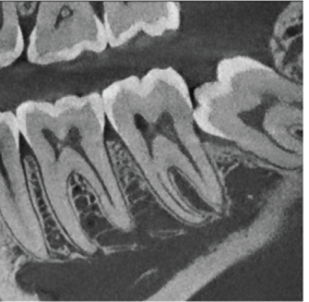
ENDO MODE - FOV 4X4
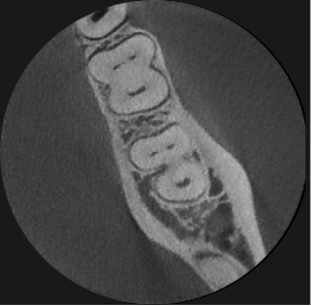
[Voxel Size :50um]
Endo TAB (Applied only on Ez3D-i Ver.5.2 on GreenX)

ENDO COLORING
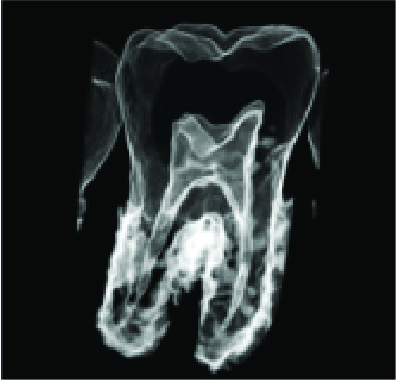
ENDO COLORING
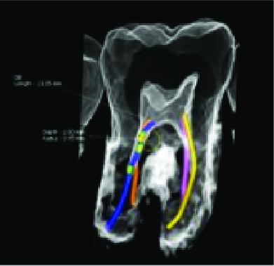
ENDO COLORING
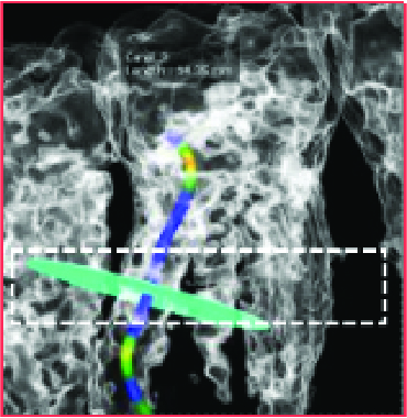
ENDO COLORING
Green X™ offers a range of selectable field of view.
The Multi FOV option allows users to select the optimum FOV mode while minimizing exposure to areas that are not in the region of interest.
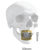
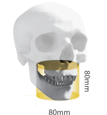
The selection includes the following FOV sizes for diagnostic needs: 16x9, 12x9, 8x8, 8x5, 5x5 and 4x4. These options cover the full arch region, sinus and left/right TMJ, and suits most oral surgery cases and multiple implant surgeries.
|
|

|

|
|---|---|---|---|
| FOV 4x4/5x5 | FOV 8x5/8x8 | FOV 12x9 | FOV 16x9 |
| - Single tooth capture | - Central dentition -TMJ (R or L) | - Dual arch including sinus and nerve -TMJ (R or L) | - Back border of jaw (Ramus) - Dual arches back to the 3" molars plus sinus - Central incisor to spine |
| - Implant single site - Endo - Perio - Complex impaction (3rd) - 0MS - Supernumerary : Ortho | - Implantology - Guided Surgery - General Dentist - 0MS - Orthodontics | - Surgical Guides - Sinus lifts - Bone grafting - Bi lateral sinus augmentation | - Surgical Guides - Sinus lifts for both sinuses - Complex orthognathic cases - Simultaneous diagnosis for both TMJs |
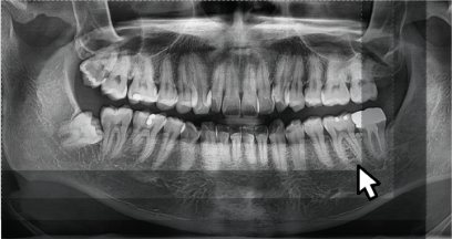
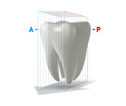
The Insight Pan is capable of taking a multilayered panoramic image, called an Insight Pan, which provides a unique in-depth look across a single focal trough.
Insight 2.0 has an upgraded free FOV feature so you will be able to capture just the area of interest.
3D model scan enables users to store plasters as digital models.
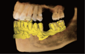
CAD/CAM integration
Sufficient level of detail for surgical guide design
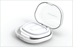
Specially Designed Jig
Stable protection from partial mode to full model
| Function | Focal Spot Size | CT FOV Size | Voxel Size | Scan Time | Gray Scale | Tube Voltage / Current | Weight | Dimensions | ||||||||||||||
|---|---|---|---|---|---|---|---|---|---|---|---|---|---|---|---|---|---|---|---|---|---|---|
| CT + Pano + Ceph + Model scan | 0.5 mm (IEC 60336) | 16x9 cm : 4x4, 5x5, 8x5, 8x8, 12x9, 16x9 cm | 4X4 0.05 mm | 5x5 0.08 mm /0.12 mm | 8x5 / 8x8 0.12 mm /0.2 mm | 12x9 / 16x9 0.2mm /0.3 mm | Pano 40sec / 14.1 sec | Ceph 1.9 sec / 4.9 sec | CBCT 2.9 sec /9.0 sec | 14 Bit | 60-99 kVp /4-16mA | Without CEPH unit 162.9 kg (359.13 lbs - without Base) | 217.9 kg (480.38 Ibs - with Base) | 187.9 kg (414.25 lbs - without Base) | With CEPH unit 242.9 kg (535.50 Ibs - with Base) | 1905.5 mm (L) x 1374.9 mm (W) x 2315.4 mm (H) - without Base | With CEPH unit 1905.5 mm (L) x 1374.9 mm (W) x 2345.4 mm (H) - with Base | 1085.0 mm (L) x 1343.5 mm (W) x 2345.4 mm (H) - with Base | Without CEPH unit 1085.0 mm (L) x 1343.5 mm (W) x 2345.4 mm (H) - with Base |
* The specifications are subject to change without prior notice.
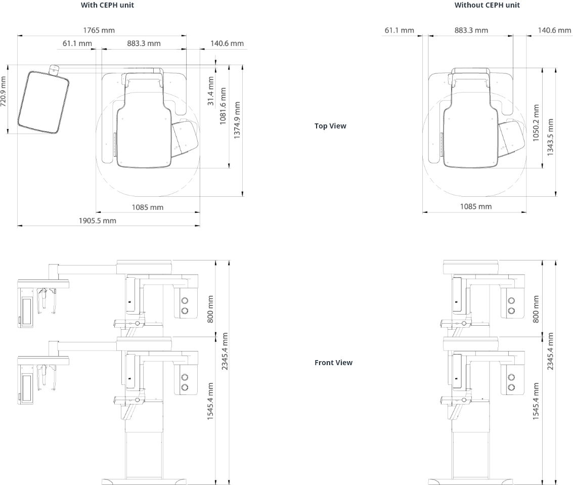
*An additional 3 inches (76.2 mm) of space is required behind the unit for wall mount bracket installation (mandatory unless there is a base mount installation).